
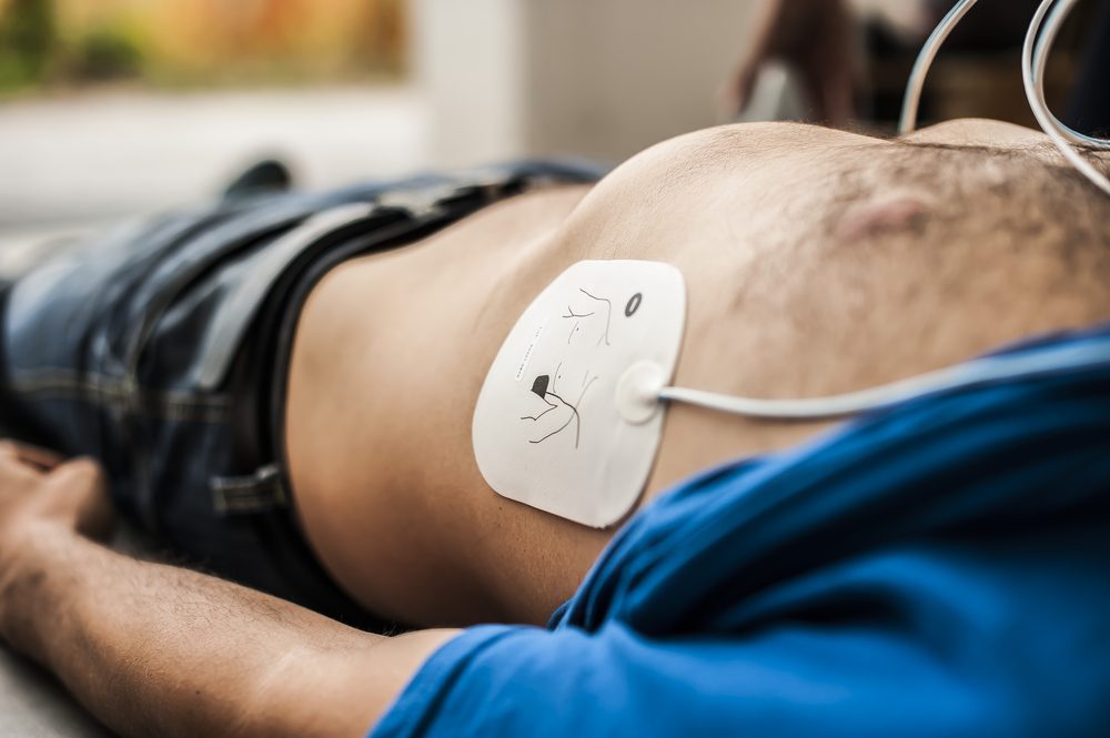
If someone arrests, do not get clever too early. High quality chest compressions, minimal interruptions, and early defibrillation for shockable rhythms still come first.
That said, some arrests will not turn around until you fix the cause. Resuscitation Council UK Adult Advanced Life Support guidance is clear on this: identify and treat reversible causes without delay. The memory aid most of us use is the 4 Hs and 4 Ts.
A quick honesty check before we go on. This is an educational overview for healthcare learners and clinically minded first aiders. It is not a substitute for an RCUK ALS course or your local protocols.
Why the mnemonic exists
Plenty of cardiac arrests, especially in hospital and in younger patients, have a treatable precipitant. RCUK's 2025 Adult ALS key points keep returning to the same themes: start ALS early, protect CPR quality, oxygenate and ventilate properly, and treat reversible causes promptly. The special circumstances guidance says the same thing in different words. Work through hypoxia, hypovolaemia, electrolyte problems, temperature extremes, tamponade, tension pneumothorax, thrombosis and toxins.
NICE Clinical Knowledge Summaries list that same family of reversible or aggravating factors. They also remind us that ischaemic heart disease remains the commonest underlying cause in many out of hospital series. So use the mnemonic as a checklist, not as a guess at every arrest.
The 4 Hs
Hypoxia
Not enough oxygen getting in. Open the airway, ventilate effectively, and correct anything reversible in the airway or breathing early. This is often the most immediately useful H.
Hypovolaemia
Not enough circulating volume for the heart to pump. Think major haemorrhage, severe dehydration, or distributive shock such as anaphylaxis or sepsis. Treat the cause and restore volume. Compressions alone will not fix an empty tank.
Hypo / hyperkalaemia and other metabolic disorders
Potassium swings and related metabolic problems can make the myocardium unstable. Hyperkalaemia is a classic issue in renal failure and some drug toxicities. If you suspect it, follow your hyperkalaemia and ALS protocols rather than improvising.
Hypo / hyperthermia
Core temperature at either extreme changes how the arrest behaves and how you manage it. In profound hypothermia, specialist pathways and careful decisions about futility matter. Do not rely on half remembered slogans.
The 4 Ts
Thrombosis (coronary or pulmonary)
Coronary occlusion is a common cause of sudden cardiac arrest. A massive pulmonary embolus can obstruct right heart outflow. ALS teaching allows you to consider fibrinolysis during CPR when PE is strongly suspected. Routine thrombolysis for suspected myocardial infarction during CPR is not supported in the same way. After ROSC, reperfusion decisions follow your local cardiac arrest centre pathway.
Tension pneumothorax
Rising pressure in the chest stops venous return. It is hard to diagnose confidently during CPR on clinical signs alone. In trained hands, ultrasound and prompt decompression (needle or finger thoracostomy, depending on competence and protocol) are part of treating the cause.
Tamponade
Fluid in the pericardial sac stops the heart filling. Think trauma, recent cardiac procedures, or known malignant disease. Ultrasound helps if you are trained to use it without wrecking your compression fraction. Drainage belongs with competent clinicians.
Toxins
Overdose, poisoning or adverse drug effects. Get specialist toxicology advice early. In the UK that usually means NPIS. Use specific antidotes where they exist, and remember that selected toxic arrests may need prolonged resuscitation under guidance.
How to use this in a real arrest
Keep CPR quality first. Do not abandon the algorithm to complete a neat list.
Give someone the job of working through the Hs and Ts while the team leader protects rhythm checks, drugs and defibrillation.
Use the history. Dialysis, asthma, trauma, pregnancy, overdose and known PE risk should push certain causes up the list.
If your team is trained for it, point of care ultrasound can support diagnosis of tamponade, pneumothorax, hypovolaemia or right heart strain. Keep pauses short.
After ROSC, keep treating the cause. Return of output is not the end of the job.
Where this sits for HealthCore learners
On Adult and Paediatric Basic Life Support, your job is still the basics: safe approach, recognition, CPR, AED use and getting help fast. The 4 Hs and 4 Ts matter more as you move into clinical roles and ALS level practice.
Our nurse led Basic Life Support (BLS) sessions build those foundations. If you need ALS, train to current RCUK standards rather than leaning on a blog post.